Overview

Hyperpigmentation is classified as solar lentigines, melasma, or post-inflammatory hyperpigmentation

Hyperpigmentation is a chronic skin condition that affects 1 in 3 people in the UK, typically those over the age of 35.

It tends to affect women more than men.

Hyperpigmentation is more common in people with darker skin tones, but it is still significant in those with lighter skin tones.

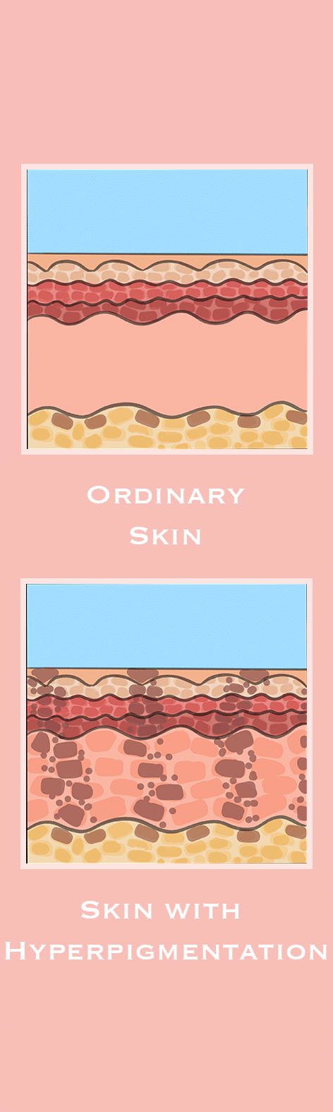
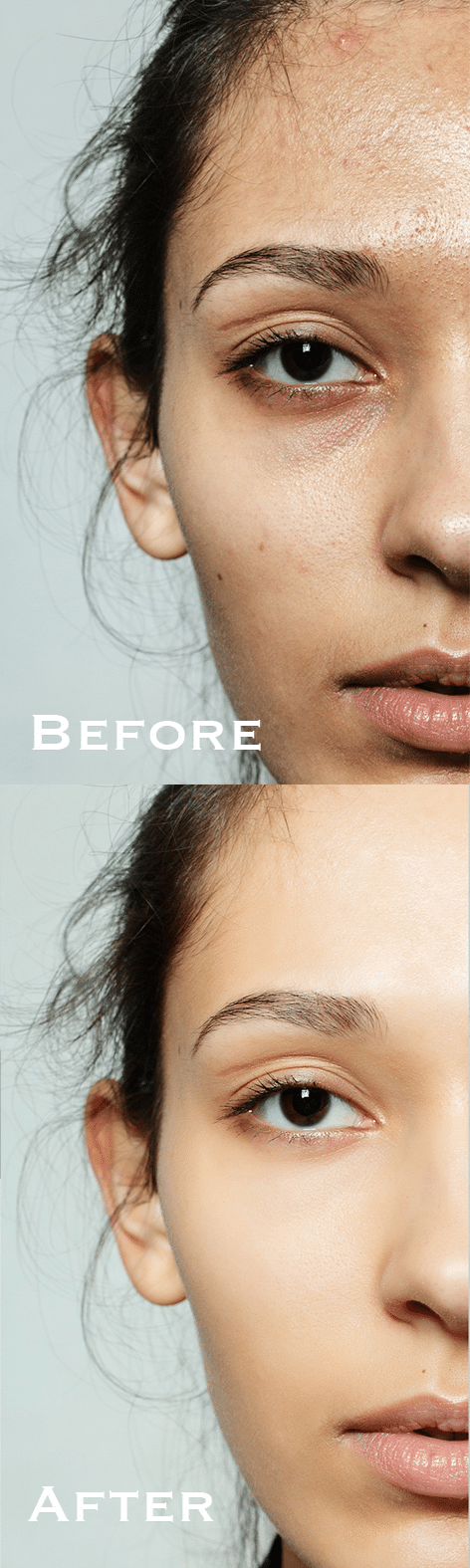
The symptoms of hyperpigmentation are patches of skin becoming darker than the surrounding skin.

The face is the most commonly affected area, accounting for 90% of all cases of hyperpigmentation. It also affects 20% of people on the neck and chest, 10% on the shoulders and back, 30% on the arms and 10% on the legs.

Hyperpigmentation consists of 3 subtypes:

Subtype Description Common Areas
Solar Lentigines

(age, liver, or sun spots)

Flat, brown, or black spots, small and can cluster, with a smooth texture Exposed areas of the skin, including the face, neck, arms, and legs
Melasma

(chloasma or the mask of pregnancy)

Flat, brown or grey-brown patches, with irregular, blotchy pigmentation, and symmetrical distribution Face, neck, stomach, and forearms
Post-Inflammatory Hyperpigmentation

(PIH)

Flat, red, brown, or black spots or patches following inflammation or injury, varying in size Any area of the body that has experienced inflammation due to a skin condition such as acne or eczema, or from an injury, like a cut or burn

Around 50% of people with hyperpigmentation develop solar lentigines, 25% melasma, and 50% post-inflammatory hyperpigmentation.

The term “hyper” refers to more, whereas “pigment” denotes colour.

Causes

Hyperpigmentation is caused by an increase in melanin production and deposition

Hyperpigmentation is caused by an overproduction of melanin, the pigment that is responsible for the colour of the hair, eyes, and skin.

The causes of hyperpigmentation include:

Cause Description
Photoageing Photoageing, caused by prolonged and unprotected exposure to ultraviolet (UV) radiation, is the leading causes of solar lentigines. While primarily resulting from sun exposure, artificial UV sources like tanning beds can contribute. Over time, cumulative UV radiation stimulates melanin production in areas most frequently exposed to the sun, such as the face, arms, and hands, leading to the formation of sun spots
Injury Injury to the skin, whether from acne, eczema, burns, or wounds, causes inflammation. As the skin heals, it can produce excess melanin, which gets deposited in the injured area, resulting in dark spots or patches that can appear red, brown, grey, or black, depending on the person’s skin tone. Further irritation, like rubbing or scratching the area, can worsen this post-inflammatory hyperpigmentation. Additionally, fungal, viral, or bacterial infections may contribute to hyperpigmentation during the healing process

Several other factors can influence the development of the condition. This includes:

Contributor Description
Genetics Genetics influence the likelihood of developing hyperpigmentation, with some people naturally more predisposed. For example, freckles are often inherited and more common in people with fair skin because of the MC1R gene. Melasma tends to run in families. Lentigines are linked to genetic conditions like LEOPARD or Peutz-Jeghers syndromes. Congenital dermal melanocytosis (CDM), often seen in some ethnic groups, is genetic. People with darker skin are more genetically predisposed to post-inflammatory hyperpigmentation because their skin cells produce more pigment
Hormones Hormones, particularly oestrogen and progesterone can cause an increase melanin, especially during pregnancy or from birth control, leading to conditions like melasma. Adrenocorticotropic hormone (ACTH) triggers melanin production, causing dark spots in conditions like Addison’s disease. Melanocyte-stimulating hormone (MSH) boosts melanin levels and can be influenced by stress or pregnancy. Thyroid imbalances, especially hypothyroidism, and insulin resistance can lead to darker patches like acanthosis nigricans
Medications Medications can increasing melanin production or depositing pigments in the skin. Antibiotics, such as tetracyclines, can cause hyperpigmentation, particularly in areas exposed to the sun. Antimalarial drugs, like chloroquine and hydroxychloroquine, can lead to blue-grey pigmentation, especially on the face. Oestrogen and progesterone found in birth control pills or hormone replacement therapy can cause melasma. Nonsteroidal anti-inflammatory drugs (NSAID’s), amiodarone, used for heart conditions, and anticonvulsants like phenytoin can result in pigmentation changes as well. Chemotherapy drugs, such as bleomycin and busulfan, are known to cause dark patches on the skin

Hyperpigmentation is common on darker skin tones, which already have a higher melanin content.

Diagnosis

Hyperpigmentation can be difficult to differentiate and diagnose, resulting in incorrect treatment and worsening of affected areas

A doctor, pharmacist, or nurse can diagnose hyperpigmentation by performing a physical examination to assess the extent and severity of the affected areas, and reviewing the patient’s medical history.

Its clinical manifestations can vary widely, and diagnosis relies on recognising characteristic patterns and understanding potential contributing factors.

The severity of hyperpigmentation can range from mild, localized discoloration to extensive, darker patches.

Severity Distribution Characteristics
Mild 1-19% Localised areas on sun-exposed areas
Moderate 20-49% Noticeable patches of darker skin, more frequent, and larger spots
Severe >50% Deep, dark patches, extensive uneven skin tone

When diagnosing hyperpigmentation, it’s important to consider other conditions that may present with similar symptoms:

Condition Description
Acanthosis Nigricans Acanthosis Nigricans manifests as dark, thick, velvety patches in skin folds like the back of the neck, armpits, and groins, and is often associated with insulin resistance, obesity, or hormonal conditions like PCOS
Addison’s Disease Addison’s Disease leads to generalised skin darkening, particularly on pressure points and skin folds, due to adrenal dysfunction, and is usually accompanied by fatigue and low blood pressure
Erythema Dyschromicum Perstans Erythema Dyschromicum Perstans is characterised by greyish or ashy patches on the face and neck, frequently linked to genetic and environmental factors, and is more common in darker skin tones
Hemochromatosis Hemochromatosis results in bronzed or darker skin due to excess iron deposits, typically accompanied by liver disease and joint pain
Lichen Planus Pigmentosus Lichen Planus Pigmentosus causes dark patches on the face and neck, is often linked to autoimmune conditions or sun exposure, and is more common in darker skin tones
Lupus Erythematosus Lupus Erythematosus often leads to dark spots following a red rash, especially on sun-exposed areas like the nose and cheeks, and is associated with joint pain and fatigue
Peutz-Jeghers Syndrome Peutz-Jeghers Syndrome presents with dark spots around the mouth and fingers, which are frequently associated with gastrointestinal polyps and an increased cancer risk
Tinea Versicolor Tinea Versicolor appears as fungal infection-induced patches of lighter or darker skin, commonly on the chest or back, affecting melanin production

A skin biopsy may be necessary to rule out another condition.

Treatment

Approaches to treating hyperpigmentation typically come in the form of topical agents or non-invasive procedures

The most effective treatment tends to include a combination of one or more of these treatments, such as a retinoid with a skin-bleaching agent.

Topical treatments can include the following:

Topical Common Examples
Retinoids Adapalene, tretinoin, and tazarotene, vitamin A derivatives, regulate the growth of cells on the skin to improve its texture and tone
Lightening Agents Hydroquinone reduces the production of melanin to lighten patches of darker skin
Anti-Inflammatories Azelaic acid inhibits the production of inflammatory-causing molecules to reduce redness and irritation
Corticosteroids Fluocinolone acetonide inhibits the production of melanin to reduce swelling and inflammation
Antioxidants Niacinamide eliminates free radicals by repairing and strengthening the barrier function of the skin, improving hydration

It is uncommon to treat hyperpigmentation orally, but emerging evidence has identified the following:

Oral Common Examples
Melanin Synthesis Inhibitors Tranexamic acid inhibits the production of melanin to reduce inflammation and can be used to treat melasma

Hyperpigmentation can be treated effectively by non-invasive procedures:

Procedure Common Examples
Exfoliation Microdermabrasion exfoliates the skin’s outermost layer using a device that sprays tiny crystals or employs a diamond-tipped wand to remove dead skin cells, stimulate cell regeneration, and promote collagen production, which helps to fade solar lentigines, melasma, and post-inflammatory hyperpigmentation
Chemical Peels Superficial peels use alpha hydroxy acids (AHA’s) like glycolic acid or beta hydroxy acids (BHA’s) like salicylic acid to exfoliate the outermost layer of skin, helping to fade minor dark spots and improve skin texture. Medium peels penetrate deeper with stronger agents such as trichloroacetic acid (TCA) or higher concentrations of glycolic acid to address more stubborn hyperpigmentation, including melasma or post-inflammatory hyperpigmentation (PIH). Deep peels utilise strong acids like high-strength TCA or phenol to reach the middle skin layers, treating severe hyperpigmentation and deeper discoloration, such as sun spots
Laser Therapy Non-ablative lasers penetrate the skin’s middle layer, stimulating collagen growth and tightening without damaging the surface, helping to reduce dark spots. Intense pulse light therapy (IPL), also known as photofacial, is a form of non-ablative, fractional laser treatment that increases collagen production, and works especially well on flat spots. Ablative lasers are the most powerful, as they remove the outer skin layers to treat deep pigmentation, causing new skin cells to grow tighter and toned

Chemical and laser peels can increase your skin’s sensitivity to sunlight.

Complications

Hyperpigmentation is a benign condition that typically does not pose health risks, however it can lead to complications that are rare, but must not be missed

People with hyperpigmentation that changes colour or does not improve with treatment should consult a doctor, chemist, or nurse. They will conduct a more thorough examination of the condition and, if necessary, perform a skin biopsy.

The complications and challenges associated with hyperpigmentation include:

Complication Description
Recurrence In cases of post-inflammatory hyperpigmentation (PIH), ongoing skin inflammation can result in pigmentation to persist and worsen over time. Even after successful treatment, hyperpigmentation may reappear if the underlying cause is not addressed and proper sun protection practices are not followed
Scarring Scarring can occur, primarily as a result of post-inflammatory hyperpigmentation. When the skin is repeatedly injured, irritated, or exposed to sunlight without protection, the risk of deeper and longer-lasting pigmentation rises, resulting in uneven skin texture and tone. In some cases, this could be more difficult to treat than the original hyperpigmentation. Acne-related hyperpigmentation may indicate deeper scarring, requiring more intensive treatment
Texture Changes Texture changes in hyperpigmentation occur when the skin becomes uneven, rough, or raised as a result of inflammation or injury. These changes often accompany dark spots, particularly when the healing process is hampered by factors like picking at wounds or sun exposure. The combination of discolouration and irregular texture can make hyperpigmentation more visible
Treatment-Related Treatment like microdermabrasion, chemical peels, and laser therapy carry risks such as scarring, infection, or worsened pigmentation if not performed correctly or without proper aftercare. Furthermore, if applied inconsistently or without professional guidance, both topical treatments and procedures can result in uneven skin tone, and allergic reactions to topical ingredients can aggravate the skin, exacerbating hyperpigmentation
Underlying Health Conditions Underlying health conditions can be associated with a variety of pigmentation changes, most notably disruptions in melanin production. Inflammatory conditions such as acne, eczema, and psoriasis can cause post-inflammatory hyperpigmentation. Hormonal imbalances, such as those seen during pregnancy or with the use of birth control, can result in melasma. Addison’s disease, a disorder of the adrenal glands, causes widespread skin darkening due to increased adrenocorticotropic hormone (ACTH). Insulin resistance in polycystic ovary syndrome (PCOS) can cause dark patches, particularly on the neck and armpits. Furthermore, certain metabolic or genetic conditions, such as hemochromatosis or Peutz-Jeghers syndrome, can cause an increase in pigmentation
Psychosocial Psychosocial impacts of hyperpigmented areas, particularly those that are prominent or widespread, can lead to feelings of self-consciousness and low self-esteem. People may experience anxiety, depression, or emotional distress as a result of how their skin appears. Some people may avoid social situations, outdoor activities, or wearing clothing that reveals the affected areas, which can reduce quality of life and self-expression

Addressing hyperpigmentation early and adhering to sun protection measures are key steps in minimising the risk of complications and improving skin appearance and overall wellbeing.

Last Updated: 10 September 2024
Next Review: 18 June 2025